Nation’s highest recognition for nursing excellence
Announcements
Please review our visitor guidelines before visiting this location. Keeping everyone safe is our top priority. We appreciate your support of our efforts.
Duke Raleigh Hospital
Open
Type:Hospital
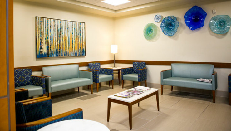
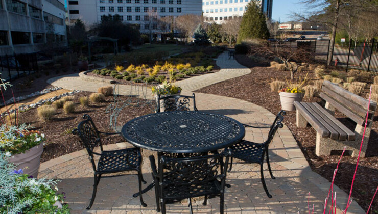
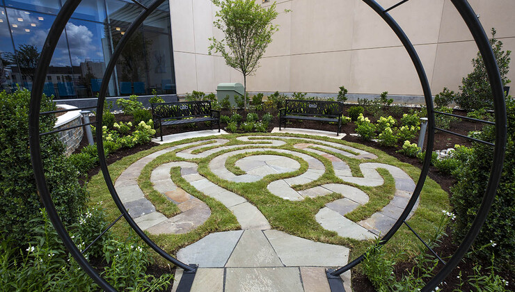
As a Duke Health hospital, we offer exceptional medical care and an outstanding experience that focuses on meeting your needs. We provide the support to help you make informed decisions about your health care.
3400 Wake Forest Rd
Raleigh,
NC
27609
General Information
919-954-3000
Connect with us:

![]()
Connect with us:
![]()
3400 Wake Forest Rd
Raleigh,
NC
27609
General Information
919-954-3000
Our Specialties
Our experienced, compassionate medical professionals deliver a comprehensive range of diagnostic, medical and surgical services in these specialties and more.
- Cancer Care
- Cardiology
- Gastroenterology
- Lung Disease
- Neurology
- Neurosurgery
- Ophthalmology
- Orthopaedic Care
- Otolaryngology
- Palliative Care
- Spine and Back Care
- Urology


Our Doctors
Find a doctor that’s right for you or your loved ones.
If you are a referring physician or health care professional, we can help you find the right people, programs, and services for your patients.